The Second Brain No One Had Seen: The Hidden Network of Astrocytes
A Reconsideration of the Role of Non-Neuronal Cells in the Central Nervous System
In classical neurophysiology, neuronal axons have always been recognized as the main mediators of functional connectivity between different brain regions. However, in recent decades, the role of glial cells—and especially astrocytes—has shifted from mere supportive cells to key players in regulating neural activities. Astrocytes communicate with each other via structures known as gap junctions: membrane channels that connect the cytoplasm of adjacent cells, enabling resource redistribution and the sharing of biochemical signals. Evidence has demonstrated that these networks are essential for memory formation, synaptic plasticity, coordination of neuronal signaling, and even the closure of critical developmental periods. Despite this significance, due to technical limitations in studying intact networks in the living organism, the spatial architecture and functional topology of these networks had remained largely unknown.
In this context, a recent article entitled Astrocytes connect specific brain regions through plastic networks: A New Paradigm in Brain Communication, by Melissa L. Cooper and colleagues, published in 2026 in the prestigious journal Nature, addresses this gap. The main objective of this research was to develop a novel method for mapping astrocytic networks throughout the brain and to investigate whether astrocytes form only a local network or possess long-range, specific communication systems akin to neuronal networks. The significance of this study lies in its provision of a tool that enable observation of astrocytic connectivity in awake, behaving animals without tissue destruction, as occurs with traditional methods such as slice electrophysiology. Prior to this study, our knowledge of astrocytic communication was limited to “dye diffusion” in acute brain slices—a method that, because of tissue damage, disrupted local connections. The question remained unanswered whether astrocytes formed a syncytium and an extensive network across the brain, or functioned as distinct sub-networks dedicated to specific regions.
It was also unclear whether the architecture of these networks aligned with neuronal pathways or provided an independent framework for non-neuronal signaling.

Innovation in Tracing: A Tool for Probing the Brain’s Hidden Networks
Researchers in this study hypothesized that astrocytes, through plastic networks based on gap junctions, connects distant brain regions in a targeted and organized manner (rather than via random diffusion), and that these networks undergo structural changes in response to environmental alterations.
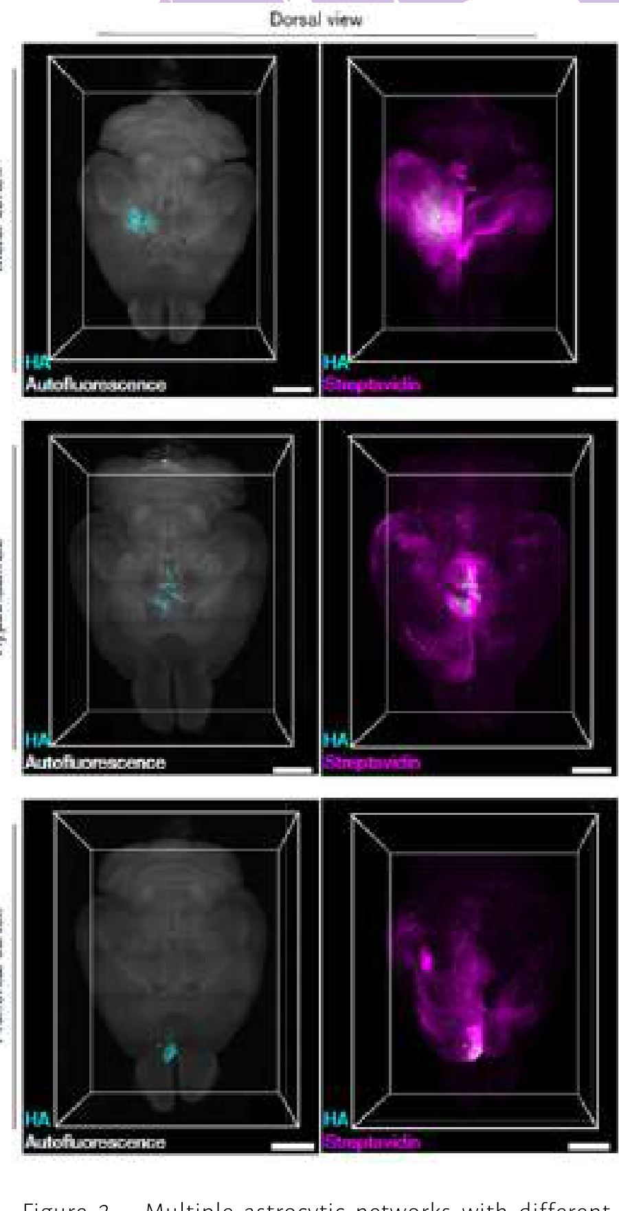
Despite the importance of astrocytic gap junction networks, studying them has always been challenging because conventional methods such as tissue slice electrophysiology disrupt network connections and, due to tissue damage, introduce experimental artifacts. To overcome these limitations, the research team devised a virus-based approach. The researchers fused the connexin protein Cx43 (the main protein forming gap junctions in astrocytes) with the enzyme TurboID (TID). As a proximity labeling enzyme, TurboID can rapidly biotinylate molecules passing through gap junction channels. Using this labeling, the movement of molecules between astrocytes connected by gap junctions became traceable (Figure 1). The study’s results showed that astrocytic networks are spread throughout the mouse brain and molecules diffuse across this interconnected network via gap junctions. However, contrary to previous assumptions, this diffusion is not random.
The researchers found that multiple networks selectively connect specific brain regions and do not pass through others. For example, the connections and network pattern of astrocytes in the motor cortex were entirely specific to that region; likewise, the hypothalamic network had a distinct set of connections and organization (Figure 2).
Some networks were restricted to a single region, but long-range networks robustly linked multiple areas across both hemispheres. This finding—that astrocytes in one hemisphere are directly networked with their counterparts in the opposite hemisphere—was among the most important results of the study.
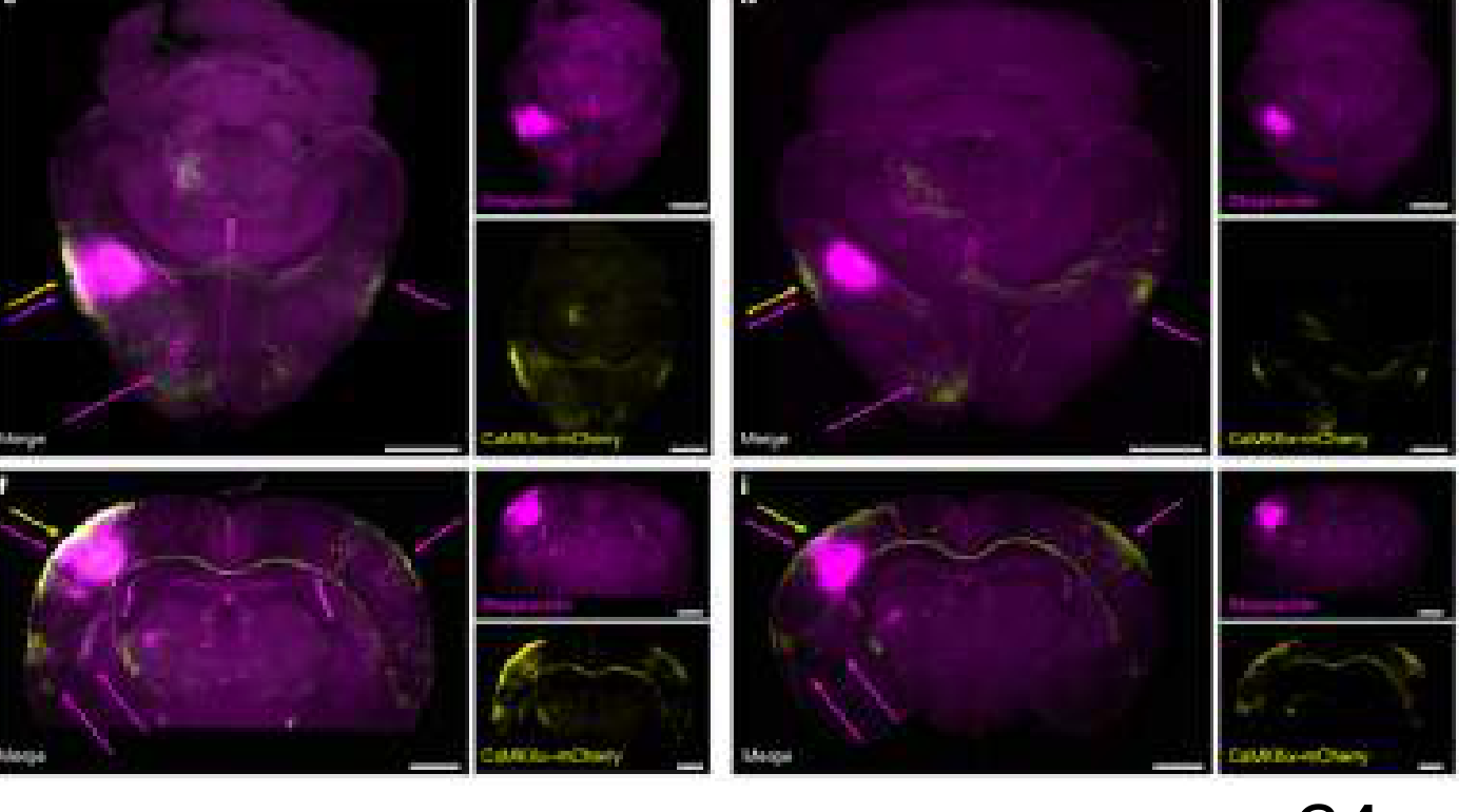
In a particularly notable part of the experiment, the researchers trimmed the whiskers of mice (Sensory deprivation) and observed that the astrocyte network in the barrel cortex became significantly smaller. This indicated that astrocytic networks, like neurons, are capable of plasticity and undergo remodeling in adulthood (Figure 3).
Although neuronal and astrocytic networks sometimes overlapped, the architecture of astrocyte networks was often distinct from known neuronal pathways, indicating an independent communication system in the brain.
The findings of Cooper et al. represent a revolution in our understanding of brain architecture; the fact that astrocytic networks can link distant regions across hemispheres shows that our brains do not contain only a “neural network,” but also a parallel, non-neuronal communication system that dynamically reorganizes in response to environmental experiences (such as sensory deprivation).
Innovations and Strengths of the Study
This study introduced several fundamental innovations:
First, the astrocytic network tracer tool made it possible for the first time to trace molecular flux—not just the physical presence of proteins.
Second, combining this method with whole-brain tissue clearing (not CLARITY, but a solvent-based technique) provided a comprehensive and three-dimensional view that was previously impossible.
Third, demonstrating glial network plasticity in response to sensory experience changed our perspective on non-neuronal cell dynamics.
Significance of Findings and Conclusion
The findings of Cooper et al. challenge the prevailing “neuron-centric” paradigm: in addition to its neural network, the brain also possesses a parallel, dynamic layer of astrocytic connectivity. These networks likely redistribute vital resources, including antioxidants like glutathione and energy sources like phosphocreatine, from less active regions to those with greater demand; moreover, biotin labeling in neuronal populations indicates that direct astrocyte-neuron communication via gap junctions may be more common than previously thought.
In the field of pathophysiology, this architecture could explain delayed or unexpected astrocyte responses in the contralateral hemisphere in diseases such as stroke and glaucoma, acting as a pathway for the spread of damaging molecules or protective signals.
If astrocytes indeed carry out this logistical role, it appears that disruption of these “hidden networks” could contribute to the progression of diseases like Alzheimer’s, Parkinson’s, and amyotrophic lateral sclerosis (ALS)—a point underscoring that our understanding of brain pathophysiology will remain incomplete without considering glial networks.
This framework opens a new avenue for neurodegenerative therapies: instead of directly targeting damaged neurons, we might bolster the brain’s “support and logistics lines.”
This raises an important question: could manipulating this plasticity protect neurons from death during inflammatory storms?
Limitations of the Study
The current approach offers a binary picture—connected or disconnected—whereas in reality, a wide spectrum of molecules crosses these networks at varying rates.
The precise temporal dynamics of molecular transfer remain unclear; the observation of certain isolated biotin-labeled astrocytes may reflect cells that were previously connected but have since become uncoupled.
The possibility that TurboID-mediated biotinylation interferes with other endogenous biotinylation pathways warrants more rigorous control experiments in future studies.
Definitively confirming the mechanism in complex genetic models remains challenging, since complete ablation of gap junctions can compromise overall cell viability.
Future Directions
Future research avenues include the use of mass spectrometry to precisely identify biotinylated molecules, in order to determine which messengers are exchanged within these networks. Investigating how brain injury, aging, or learning reshape the architecture of these networks could also open new therapeutic targets in neurology.
This article is adapted from the following study: