The history of modern neuroscience began with a profound paradox. The brain, recognized today as one of the most complex biological structures known to science, appeared under the microscopes of the nineteenth century as little more than a homogeneous, gelatinous mass devoid of clearly distinguishable boundaries. Today, it is self-evident that the cerebral cortex is composed of billions of neurons interconnected through an immense network of processes. However, until the 1870s, the limited resolution of techniques available for visualizing the fine architecture of neural tissue prevented scientists from appreciating its precise structural organization [1].
By that time, fixation and staining techniques in general anatomy had undergone considerable development. Scientists could readily distinguish hepatic cells and muscle fibers using natural dyes such as carmine and hematoxylin. Neuroanatomy, however, remained at a major impasse.
The brain presented two particularly challenging characteristics that rendered the histological tools of the era largely ineffective:
- The extraordinarily high density and extensive intermingling of cellular processes, collectively forming the neuropil.
- Its high lipid content, particularly the sphingolipids and cholesterol-rich membranes of myelin.
The commonly used dyes of the time either failed to penetrate neural tissue effectively because of its hydrophobic nature or stained virtually all cellular structures simultaneously and uniformly. The resulting microscopic image was therefore obscure and poorly resolved—much like a dense rainforest in which the boundaries of an individual tree cannot be distinguished from those of neighboring trees because of the overwhelming density of foliage. Prominent anatomists such as Johannes Purkinje and Otto Deiters attempted to overcome this problem by producing extremely thin sections with very sharp blades, thereby physically reducing tissue density; nevertheless, cellular boundaries remained indistinct [1].
When Neurons Were Seen for the First Time
In 1873, this historical impasse was broken by Camillo Golgi, a young physician who, owing to financial constraints, had been forced to abandon his academic position and accept the superintendence of a hospital for chronically ill patients in the small town of Abbiategrasso. He established a rudimentary laboratory in the kitchen of his residence, where he developed a new protocol that would later become known as the Golgi stain or Golgi impregnation method [2].
In this technique, Golgi initially fixed brain tissue in a solution of potassium dichromate. This biochemical step not only increased tissue rigidity but also prepared the tissue for the subsequent penetration of ions into cellular structures. The fixed tissue was then transferred into a solution of silver nitrate. At this stage, silver ions reacted with the chromate ions that had penetrated the tissue, resulting in the formation of silver chromate crystals. These dark brown or black precipitates accumulated within the neuronal soma and cellular processes [2].
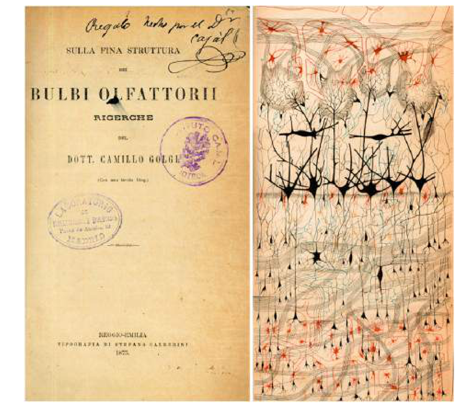
DeFelipe J. Cajal and the discovery of the Golgi method: a neuroanatomist’s dream. Anat Sci Int. 2025;100(4):384–399. doi:10.1007/s12565-025-00840-7.
The Clash of Two Perspectives and the Ascent to the Peak of the World’s Greatest Scientific Honor
This powerful morphological tool reached Santiago Ramón y Cajal, the patient Spanish anatomist, in 1887. Cajal recognized several limitations of Golgi’s original method: silver deposits could sometimes be unstable, while the extensive presence of myelin in mature neural tissue interfered with the precise tracing of axonal trajectories [3].
Cajal introduced two strategic modifications to the protocol:
- First, double impregnation: he repeated the fixation and silver-impregnation process, increasing the density of the deposits and revealing delicate structures such as dendritic spines.
- Second, adoption of an ontogenetic approach (examining the brain during early developmental stages): rather than focusing primarily on the adult brain, which possesses a more complex neural network and abundant myelination, Cajal examined embryonic brains and those of newborn animals. At these developmental stages, the neural network is less densely packed and myelination has not yet become extensive. This relative structural simplicity allowed him to trace the trajectory of individual nerve fibers (axons) with unprecedented clarity [3].
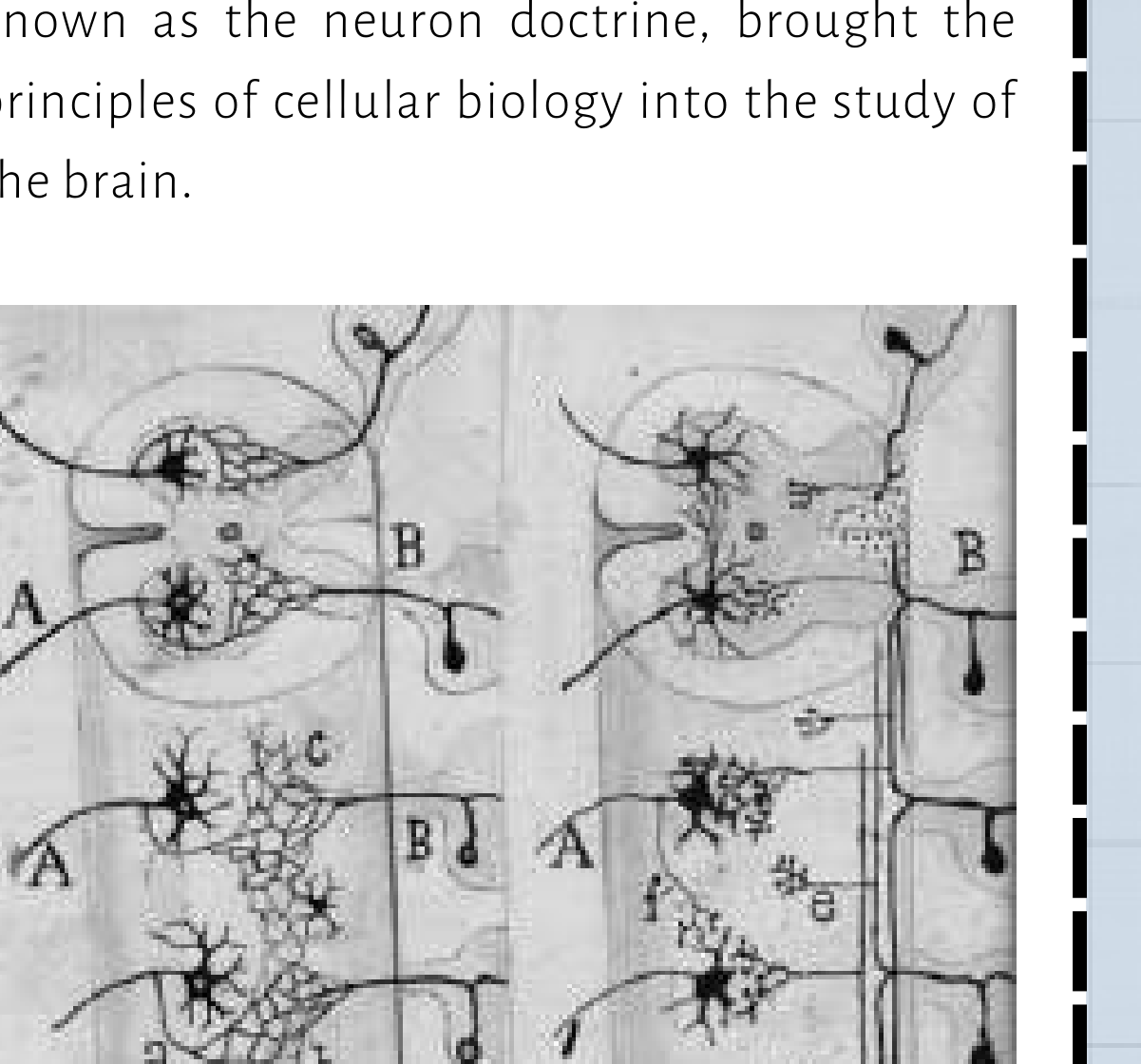
This technical optimization provided the spark for one of the greatest intellectual confrontations in the history of science. Based on his observations of Golgi-stained preparations, Camillo Golgi adhered to the reticular theory, arguing that the nervous system constituted a continuous cytoplasmic network, or syncytium, analogous in some respects to the vascular system [2]. In contrast, drawing on his improved preparations, Cajal observed the physical individuality of individual cells and demonstrated that neurons are discrete structural and functional units that communicate through specialized contacts across microscopic gaps—the synaptic clefts. This concept, which became known as the neuron doctrine, brought the principles of cellular biology into the study of the brain.
The disagreement was so profound that even when the two scientists jointly received the 1906 Nobel Prize in Physiology or Medicine, Golgi used his Nobel lecture to characterize the neuron doctrine as an outdated hypothesis. The following day, Cajal defended his hypothesis by presenting compelling anatomical evidence in support of neuronal individuality [4].
The Legacy of the Industrial Revolution in Mapping the Cerebral Cortex: How Specialized Stains Emerged
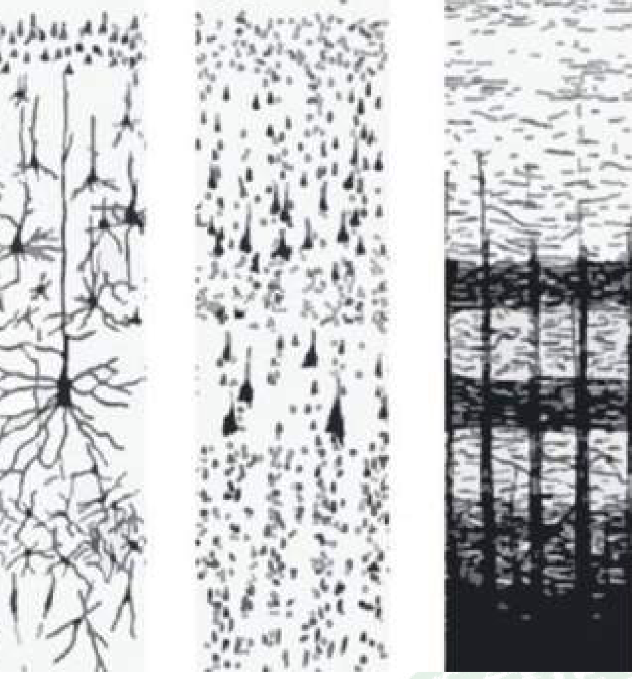
Despite the effectiveness of the Golgi method for examining the morphology of individual neurons, its sparse and selective staining pattern prevented it from revealing the overall organization and laminar architecture of the brain. By the late nineteenth century, Germany had established dominance over the emerging industry of synthetic organic dyes, particularly aniline dyes derived from coal tar. This industrial capacity helped transform German universities into leading centers of histological research.
During this period, Franz Nissl discovered that basophilic dyes such as cresyl violet exhibited a strong electrostatic affinity for acidic intracellular components, particularly nucleic acids and, most notably, ribosomal RNA. In neurons, the structures that became known as Nissl bodies corresponded primarily to the densely packed rough endoplasmic reticulum within the soma and proximal dendrites. Because axons lack rough endoplasmic reticulum, they remained unstained by the Nissl method. This distinction enabled Korbinian Brodmann, through systematic examination of the cellular architecture of the cerebral cortex, to construct his famous map of 52 cortical areas [1]. At approximately the same time, Carl Weigert developed a staining method using hematoxylin modified with copper salts that preferentially targeted the lipids of the myelin sheath. In contrast to Nissl staining, this technique highlighted the brain’s communication cables—the white matter—by darkening myelinated fiber tracts and thereby revealing the brain’s major pathways of connectivity [1].
Rereading the Brain’s Identity with Fluorescent Neurons
By the second half of the twentieth century, classical histology had encountered a major limitation: these staining methods could not provide functional or molecular specificity. To conventional histological stains, a dopaminergic neuron was essentially indistinguishable from a serotonergic neuron. To overcome this limitation, scientists developed immunohistochemistry. Fluorescently labeled antibodies were employed as highly specific molecular probes capable of binding to proteins characteristic of particular cell types, thereby enabling chemical and molecular mapping of the brain [6].
But the next major question was this: What if the neuron itself could emit light without requiring an externally applied dye?
The answer was hidden in the genome of a hydrozoan jellyfish that naturally produces green fluorescent protein (GFP). In a revolution in genetic engineering, scientists transferred the genetic sequence encoding this protein into the genomes of laboratory animals, enabling neurons within a living brain to produce fluorescent proteins themselves [7].
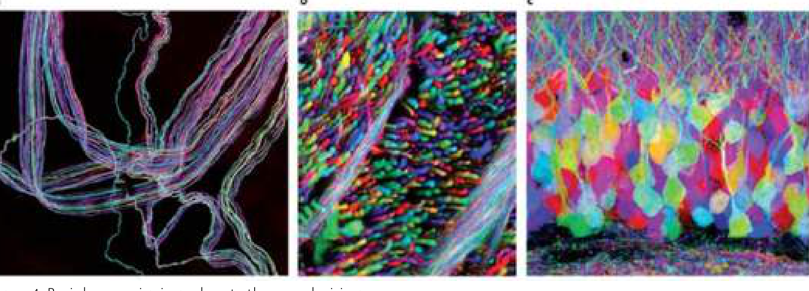
However, in the densely packed environment of the brain, when millions of neurons all emitted the same green fluorescence, distinguishing individual neuronal processes remained difficult. In 2007, researchers at Harvard University addressed this problem by introducing the technique known as Brainbow. Using stochastic genetic recombination, they expressed genes encoding multiple fluorescent proteins in the genomes of mice. Random combinations of these fluorescent proteins within individual cells—analogous to the mixing of colored pixels in a display—caused each neuron to acquire a distinctive and essentially unique color signature. The technique was therefore analogous to color-coding the wiring of a supercomputer, allowing researchers to trace individual “wires,” or neuronal processes, through thousands of neighboring fibers. It became possible to follow a purple neuronal process through a dense forest of neighboring cyan and orange branches [8].
Journey into the Glass Brain: CLARITY and Three-Dimensional Resolution
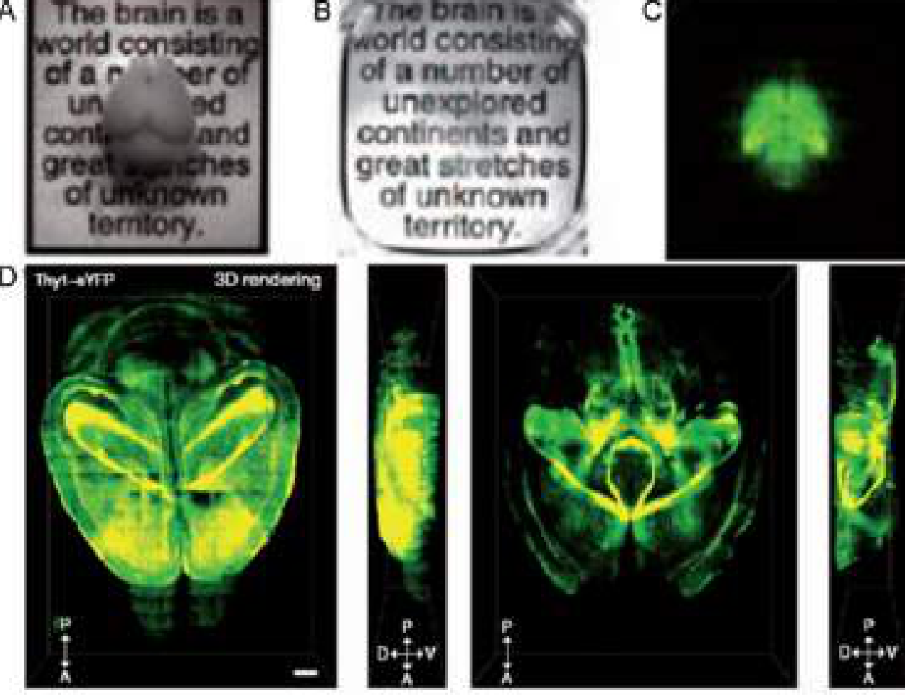
The final major obstacle was the intrinsic opacity of the brain, largely attributable to its high lipid content, particularly myelin, which interferes with the transmission of light through tissue. For many years, the only practical way to visualize structures deep within neural tissue was to mechanically section the brain, a process that inevitably disrupted long-range anatomical connections. In 2013, the remarkable CLARITY technique overcame this limitation. In this approach, brain tissue is first embedded within a polymerized hydrogel matrix that forms a physical scaffold for preserving proteins and nucleic acids. Subsequently, light-scattering lipids are removed from the tissue using detergents, facilitated by an applied electrical field. The result is a highly transparent, glass-like brain in which the three-dimensional architecture of neuronal structures can be preserved while permitting deep-tissue imaging [5].
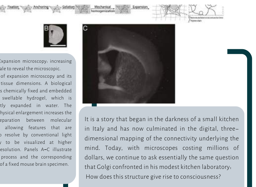
Almost simultaneously, an inverse strategy known as expansion microscopy was introduced. Rather than attempting to force light through densely packed tissue, this approach embeds the specimen within a highly water-absorbing hydrogel and induces uniform physical expansion in all directions. This expansion increases the physical distance between target molecules, enabling structures that were previously below the diffraction-limited resolution of conventional light microscopy—approximately 200 nm—to be visualized with substantially greater spatial resolution [9].
The Brain’s Fabric Through Time: From Classical Histological Staining to Digital Connectomics
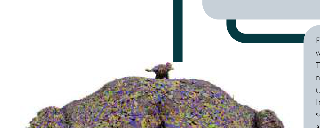
Today, large-scale connectomics projects are attempting to reconstruct comprehensive maps of the brain’s synaptic connectivity. For example, the international FlyWire consortium has successfully reconstructed the complete neuronal connectivity of the adult fruit-fly brain, while the MICrONS project has reconstructed an entire cubic millimeter of mouse cerebral cortex at synaptic resolution. These achievements were recognized by the prestigious journal Nature Methods as the method of the year in neuroscience [10].
Despite relying on sophisticated machine-learning algorithms and advanced electron microscopy, these projects remain grounded in the same conceptual principle that Cajal established with a paintbrush and a relatively simple microscope: creating contrast within densely packed biological structures in order to make the invisible visible.
It is a story that began in the darkness of a small kitchen in Italy and has now culminated in the digital, three-dimensional mapping of the connectivity underlying the mind. Today, with microscopes costing millions of dollars, we continue to ask essentially the same question that Golgi confronted in his modest kitchen laboratory: How does this structure give rise to consciousness?
References
- Shepherd, G. M. (1991). Foundations of the Neuron Doctrine. Oxford University Press.
- Golgi, C. (1873). Sulla struttura della sostanza grigia del cervello. Gazzetta Medica Italiana, 6, 244-246.
- Ramón y Cajal, S. (1911). Histology of the Nervous System of Man and Vertebrates. Oxford University Press.
- Grant, G. (2007). The Nobel Prize in Physiology or Medicine 1906: Camillo Golgi and Santiago Ramón y Cajal. Journal of the History of the Neurosciences, 16(1-2), 1-4.
- Chung, K., & Deisseroth, K. (2013). CLARITY for mapping the nervous system. Nature Methods, 10(6), 508-513.
- Coons, A. H., Creech, H. J., & Jones, R. N. (1941). Immunological properties of an antibody containing a fluorescent group. Proceedings of the Society for Experimental Biology and Medicine, 47, 200-202.
- Chalfie, M., Tu, Y., Euskirchen, G., Ward, W. W., & Prasher, D. C. (1994). Green fluorescent protein as a marker for gene expression. Science, 263(5148), 802-805.
- Livet, J., Weissman, T. A., Kang, H., et al. (2007). Transgenic strategies for combinatorial expression of fluorescent proteins in the nervous system. Nature, 450, 56-62.
- Chen, F., Tillberg, P. W., & Boyden, E. S. (2015). Expansion microscopy. Science, 347(6221), 543-548.
- Nature Methods Editorial. (2025). Method of the Year: electron microscopy-based connectomics. Nature Methods, 22.